Back Muscles Anatomy Labeled / Deep Muscles of the Back - See back muscles and low back pain nerves in your lower back five pairs of lumbar spinal nerves labeled l1 to l5 branch off your spinal cord and exit through small holes between the vertebrae.
Back Muscles Anatomy Labeled / Deep Muscles of the Back - See back muscles and low back pain nerves in your lower back five pairs of lumbar spinal nerves labeled l1 to l5 branch off your spinal cord and exit through small holes between the vertebrae.. For more anatomy content please follow us and visit our website: On this page, you'll learn about each of these muscles, their locations and functional anatomy. This website uses cookies to improve your experience while you navigate through the website. The back's muscles start at the top of the back (named the cervical vertebrae) and go to the tailbone (also named the coccyx). We are pleased to provide you with the picture named muscles of lower back diagram.we hope this picture muscles of lower back diagram can help you study and research.
The deep muscles develop embryologically in the back, and are thus described as intrinsic muscles. Human anatomy pictures of lower back muscles and human anatomy. Learn vocabulary, terms, and more with flashcards, games, and other study tools. Several small muscles in the cervical area of the vertebral column are also important. Related posts of muscles labeled front and back.
Rezultat imagine pentru leg muscle model labeled | Muscle ... from i.pinimg.com This long muscle travels nearly the entire length of the back. There is a printable worksheet available for download here so you can take the quiz with. Superficial back muscles, intermediate back muscles and intrinsic back muscles.the intrinsic muscles are named as such because their embryological development begins in the back, oppose to the superficial and intermediate back muscles which develop elsewhere and are therefore classed as extrinsic muscles. The superficial group, the deep group, and the intermediate group. Claim your free copy of the client back care guide today. Each type of muscle tissue in the human body has a unique structure and a specific role. See back muscles and low back pain nerves in your lower back five pairs of lumbar spinal nerves labeled l1 to l5 branch off your spinal cord and exit through small holes between the vertebrae. The back's muscles start at the top of the back (named the cervical vertebrae) and go to the tailbone (also named the coccyx).
Superficial back muscles, intermediate back muscles and intrinsic back muscles.the intrinsic muscles are named as such because their embryological development begins in the back, oppose to the superficial and intermediate back muscles which develop elsewhere and are therefore classed as extrinsic muscles.
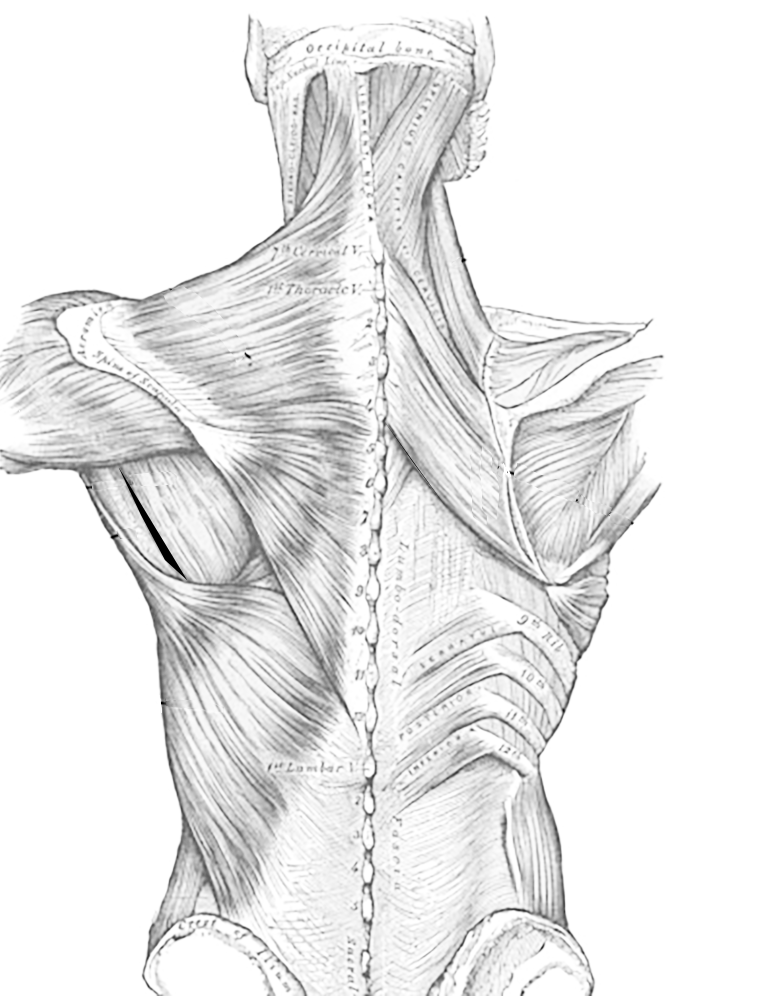
We hope this picture anatomy of back muscles diagram can help you study and research. For more anatomy content please follow us and visit our website: There is a printable worksheet available for download here so you can take the quiz with. These muscles lie on each side of the vertebral column , deep to the thoracolumbar fascia . Just need a glimpse, leave your valuable advice let us know , and subscribe us! Several small muscles in the cervical area of the vertebral column are also important. Link to client back care guide Female reproductive organs front view. The muscles of the back can be arranged into 3 categories based on their location: This long muscle travels nearly the entire length of the back. Superficial back muscles, intermediate back muscles and intrinsic back muscles.the intrinsic muscles are named as such because their embryological development begins in the back, oppose to the superficial and intermediate back muscles which develop elsewhere and are therefore classed as extrinsic muscles. This website uses cookies to improve your experience while you navigate through the website. The back consists of the spine, spinal cord, muscles, ligaments, and nerves.
These structures work together to support the body, enable a range of movements, and send messages from the brain to the. Just need a glimpse, leave your valuable advice let us know , and subscribe us! For more anatomy content please follow us and visit our website: These muscles include the large paired muscles in the lower back called erector spinae which help hold up the spine and gluteal muscles. The spine's four sections, from top to bottom, are the cervical (neck), thoracic (abdomen,) lumbar (lower back), and sacral (toward tailbone).
Muscles of the Back and Chest from www.biologycorner.com Several small muscles in the cervical area of the vertebral column are also important. Each type of muscle tissue in the human body has a unique structure and a specific role. Back muscles diagram body muscles labeled science of anatomy. The superficial and intermediate muscles do not develop in the back, and are classified as extrinsic muscles. The back consists of the spine, spinal cord, muscles, ligaments, and nerves. Human anatomy for muscle, reproductive, and skeleton. This website uses cookies to improve your experience while you navigate through the website. Muscles found in the superficial group include rhomboid major, rhomboid minor, levator scapulae, trapezius, latissimus dorsi.
Learn anatomy back labeling with free interactive flashcards.
Human anatomy pictures of lower back muscles and human anatomy. Muscle anatomy front 12 photos of the muscle anatomy front front leg muscle anatomy, front neck muscle anatomy, front shoulder muscle anatomy, frontalis muscle anatomy, muscle anatomy front, human muscles, front leg muscle anatomy, front neck muscle anatomy, front shoulder muscle anatomy, frontalis muscle anatomy, muscle. This article covers the anatomy of the deep muscles of the back, including their function, blood supply, innervation, origin and insertion. Back anatomy muscle have a graphic associated with the other. 12 photos of the muscle diagram for chest and back. Superficial, intermediate, deep and deepest layers. Back muscles diagram body muscles labeled science of anatomy. Each type of muscle tissue in the human body has a unique structure and a specific role. We hope this picture anatomy of back muscles diagram can help you study and research. Human anatomy for muscle, reproductive, and skeleton. We are pleased to provide you with the picture named muscles of lower back diagram.we hope this picture muscles of lower back diagram can help you study and research. Your clients will thank you for it! Because the lower back is relatively mobile yet carries the most load, it is the most likely area of the spine to wear down or sustain an injury.
Labeled anatomy chart of male triceps and back muscles on white background labeled human anatomy diagram of man's arm, shoulder and upper back muscles in a posterior view on a white background. There are three different muscle groups found in the back: Choose from 500 different sets of anatomy back labeling flashcards on quizlet. For more anatomy content please follow us and visit our website: The part of the nerve that emerges out of the spine is called the nerve root.
leg model - labeled muscles | Anatomy lab 2 | Pinterest ... from s-media-cache-ak0.pinimg.com Other muscles are small and cover much less space. Studying these is an ideal first step before moving onto the more advanced practices of muscle labeling and quizzes. Because the lower back is relatively mobile yet carries the most load, it is the most likely area of the spine to wear down or sustain an injury. These muscles include the large paired muscles in the lower back called erector spinae which help hold up the spine and gluteal muscles. Anatomynote.com found anatomy of back muscles diagram from plenty of anatomical pictures on the internet. Muscles of the torso labeled / extrinsic back muscles anatomy online medical library by author mei 14, 2021 third, the muscles of the torso do not move just the torso (vertebral column and rib cage) but also the shoulder girdle, which includes the there are many ways to categorize the torso muscles. The back's muscles start at the top of the back (named the cervical vertebrae) and go to the tailbone (also named the coccyx). Back muscles diagram body muscles labeled science of anatomy.
Lunges that relieve back pain low back pain program.
Some of these muscles are quite large and cover broad areas. Start studying 08 back muscles label. Superficial, intermediate, deep and deepest layers. The muscles of the chest and upper back occupy the thoracic region of the body inferior to the neck and superior to the abdominal region and include the muscles of the shoulders. 12 photos of the muscle diagram for chest and back. This long muscle travels nearly the entire length of the back. The superficial group, the deep group, and the intermediate group. These muscles include the large paired muscles in the lower back called erector spinae which help hold up the spine and gluteal muscles. Muscles of the torso labeled / extrinsic back muscles anatomy online medical library by author mei 14, 2021 third, the muscles of the torso do not move just the torso (vertebral column and rib cage) but also the shoulder girdle, which includes the there are many ways to categorize the torso muscles. Learn vocabulary, terms, and more with flashcards, games, and other study tools. These muscles lie on each side of the vertebral column , deep to the thoracolumbar fascia . The back consists of the spine, spinal cord, muscles, ligaments, and nerves. Several small muscles in the cervical area of the vertebral column are also important.
Lower back pain and sciatica back muscles anatomy. The intrinsic back muscles, which are also called true back muscles.


0 Komentar